



Biomolecular markers of inflammation in cancer
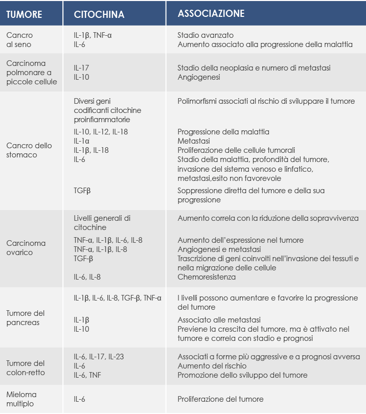
Several biomolecular markers of inflammation are also associated with cancer. One of the most widely known is C-reactive protein (CRP), a molecule whose production is regulated by pro-inflammatory cytokines. CRP levels are very low in healthy people but rise rapidly with inflammation, and they are associated with tumor progression and reduced survival in several cancers, for example of the esophagus, stomach, colon-rectum, liver, pancreas and ovary.
In general, higher CRP concentrations are associated with a greater risk of developing cancer. Pre-diagnostic CRP levels, for example, are associated with the incidence of colorectal cancer and with the risk of breast cancer. What is more, in colorectal cancer, CRP concentrations before diagnosis also correlate with cancer mortality.
As for cytokines, breast cancer offers an example of their involvement. They can be produced by inflammatory cells, by tumor cells and by the cells in the surrounding environment. High levels of interleukin 1β (IL-1β) are associated with more advanced disease, and high levels of interleukin 6 (IL-6) appear to contribute to disease progression. In general, the production of pro-inflammatory cytokines such as Tumor Necrosis Factor α (TNF-α) by tumor-associated macrophages (TAMs) may contribute to their ability to promote tumor development. Blood concentrations of TNF-α have themselves been associated with more advanced tumor stages.
In fact, among the various cytokines, TNF is a particularly important factor in promoting the development of several types of cancer. Produced in the early phase of the inflammatory response, it plays a key role in maintaining chronic inflammation and is involved in tumor growth, angiogenesis, tissue remodeling and the formation of metastases.
When metastases are present, interleukin 17 (IL-17) also rises. Together with interleukin 23 (IL-23), IL-17 expression has been found in tumors of the colon, ovary, lung, breast, stomach, skin, liver, and head and neck. In small cell lung cancer it has also been associated with the stage of the neoplasm and the number of metastases; for this reason it is thought that it could represent a new prognostic marker.
Another potent inducer of angiogenesis in small cell lung carcinoma is interleukin 10 (IL-10), which in stomach cancer instead appears to contribute to disease progression by suppressing antitumor immunity. Also in stomach cancer, an effect similar to that of IL-10 may be attributed to interleukin 12 (IL-12) and interleukin 18 (IL-18), while interleukin 1α (IL-1α) correlates with metastases, IL-1β and IL-18 stimulate the proliferation of tumor cells, and IL-6 is associated with the stage of the disease, the depth of the tumor, invasion of the lymphatic and venous systems, metastases and an unfavorable outcome. Finally, Transforming Growth Factor β (TGFβ) appears to play a complex role in the direct suppression of the tumor and of its progression (probably owing to its immunosuppressive and anti-inflammatory properties), which may depend on the stage of the disease and on the type of cells involved.
Biomolecular markers of inflammation in cancer
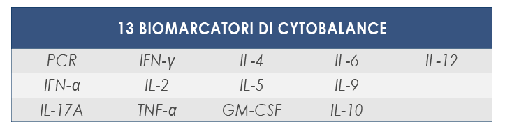
Low-grade chronic inflammation is not accompanied by the classic symptoms of acute inflammation (swelling, pain, redness and heat), but fortunately today numerous biomolecular markers make it possible to recognize its presence through a simple blood test. These include several cytokines, mediators of inflammatory processes such as TNF-α, interleukins 1, 6 and 12 and interferon, and C-reactive protein (CRP), a protein produced by the liver precisely in response to the presence of pro-inflammatory cytokines.
Pro-inflammatory cytokines
Produced by activated inflammatory cells, pro-inflammatory cytokines are regulated by several mechanisms. Among the most important is the activity of SOCS (Suppressor Of Cytokine Signaling), a protein that promotes further activation of inflammatory cells by acting on transcription factors such as NF-kB (a key regulator of inflammatory processes) and STAT (Signal Transducers and Activators of Transcription) 1 and 3.
When activated by pro-inflammatory stimuli, NF-kB leads to the coordinated expression of several genes with a pro-inflammatory action or associated with innate immunity, including IL-1, 4 and 6 and TNF-α. Many of these genes are also activated by pro-inflammatory stimuli acting on STAT1 (which is essential for the action of IFN-γ and promotes NF-kB-induced programmed cell death) and STAT3 (which, when activated by IL-10, instead promotes cell proliferation by suppressing the activity of NF-kB).
The Granulocyte-Macrophage
Colony-Stimulating Factor (GM-CSF)
Granulocyte-Macrophage Colony-Stimulating Factor (GM-CSF) is a cytokine able to stimulate the production of blood cells in the bone marrow, including the white blood cells of the granulocyte and macrophage classes. Compared with other members of the CSF superfamily, however, its role in supporting these cells is marginal, especially when compared with its role as a mediator of inflammation.
Its expression is associated with several inflammatory phenomena. It is, for example, activated in the cerebrospinal fluid of patients with multiple sclerosis; it is also activated in inflamed tissues such as the skin affected by skin reactions and the joints affected by rheumatoid arthritis, and it rises rapidly and significantly in response to lipopolysaccharide (LPS), a toxin of bacterial origin.
It is produced by several types of cells. Classically, the GM-CSF associated with chronic inflammatory diseases of autoimmune origin such as rheumatoid arthritis is synthesized by T lymphocytes following stimulation by one or more interleukins (IL-2, IL-7, or a combination of IL-1 and IL-23). In multiple sclerosis, however, it is also produced by B lymphocytes.
The tissue damage associated with the chronic inflammation promoted by GM-CSF is not, however, caused directly by lymphocytes. Rather, it would be the ability of this cytokine to link T lymphocytes with cells such as macrophages and granulocytes that triggers effects that, in chronic inflammation, can end up damaging otherwise healthy tissues, for example the production of reactive oxygen species.
Bringing elevated GM-CSF levels back to normal could help fight chronic inflammation. This approach has already proved potentially effective in people with rheumatoid arthritis. Trials carried out in multiple sclerosis have shown no unexpected side effects.
Tumor Necrosis Factor α (TNF-α) is a pro-inflammatory cytokine that is essential for maintaining low-grade systemic inflammation. Its role is indispensable, for example, in trauma or infections; however, when its levels remain constantly high a state of chronic inflammation sets in that can damage tissues. In particular, TNF-α can contribute to the malfunction of blood vessels and to the development of hypertension and atherosclerosis.
In apparently healthy individuals it is associated with blood pressure: a constant increase in its production, even at low levels, favors vasoconstriction and, as a consequence, induces hypertension. In particular, this cytokine impairs the endothelium-dependent vasodilation process (the endothelium being the epithelium lining the inside of blood vessels) by reducing the bioavailability of the main mediator of this phenomenon, nitric oxide (NO), whose production it lowers while at the same time increasing its clearance.
But that is not all: TNF-α mediates insulin resistance in blood vessels, contributing to generalized endothelial dysfunction in both diabetes and cardiometabolic syndrome. It also regulates blood pressure in the kidneys and in the heart muscle.
Also in the blood vessels, TNF-α stimulates the production of reactive oxygen species and induces the expression of adhesion molecules that mediate the recruitment and migration of inflammatory cells into the vessel wall, thus favoring the process of atherosclerosis. At the same time, it induces proatherogenic conditions by favoring the proliferation and migration of the smooth muscle cells associated with the endothelium.
Finally, it reduces the contractility of cardiac muscle cells, induces their hypertrophy and, in vitro, promotes their programmed death.
Interleukin 6 (IL-6) is a cytokine able to stimulate the acute inflammation associated with infections and tissue damage. Its production acts as an alarm and defense signal; however, when its activity remains constantly elevated the inflammatory response becomes chronic, with the possible dangers that follow. What is more, continuous production of IL-6 is involved in autoimmunity.
After being produced, IL-6 travels through the bloodstream to the liver, where several proteins associated with the acute phase of inflammation are rapidly produced, such as C-reactive protein
(CRP) and serum amyloid A (SAA). If its concentration remains high for a long time, SAA induces the deposition of amyloid fibrils, a serious complication of several chronic inflammatory diseases that leads to progressive deterioration of the organs.
In addition, IL-6 lowers blood levels of iron and zinc, leading to forms of anemia and to iron and zinc deficiency associated with chronic inflammation. And the consequences of its activation do not end there; among others they include:
- the release of platelets from the bone marrow, a marker of the severity of inflammation;
- a modulation of lymphocyte differentiation that leads to impairment of the immunological tolerance involved in the development of chronic inflammatory and autoimmune diseases;
- excessive production of immunoglobulins and synthesis of autoantibodies;
- bone resorption and osteoporosis;
- increased angiogenesis and blood vessel permeability, two pathological features of the inflammatory lesions associated, for example, with rheumatoid arthritis;
- the proliferation of keratinocytes and the production of collagen in dermal fibroblasts, associated with systemic sclerosis.
Several inflammatory or autoimmune diseases, as well as various forms of cancer, have been associated with abnormal production of IL-6. Studies in animal models have shown that blocking its action can limit or suppress the development of these diseases.
The interleukin (IL) 17 family is a group of pro-inflammatory cytokines made up of 6 distinct members. Among them, interleukin 17A (IL-17A) is produced in large amounts in several chronic diseases, such as rheumatoid arthritis, multiple sclerosis, inflammatory bowel diseases and psoriasis; its receptor (IL-17RA) is expressed in many types of cells and tissues.
Cytokines belonging to the IL-17 family can be produced by several cells of the immune system, but their main source is a specific type of T lymphocytes, known as Th17.
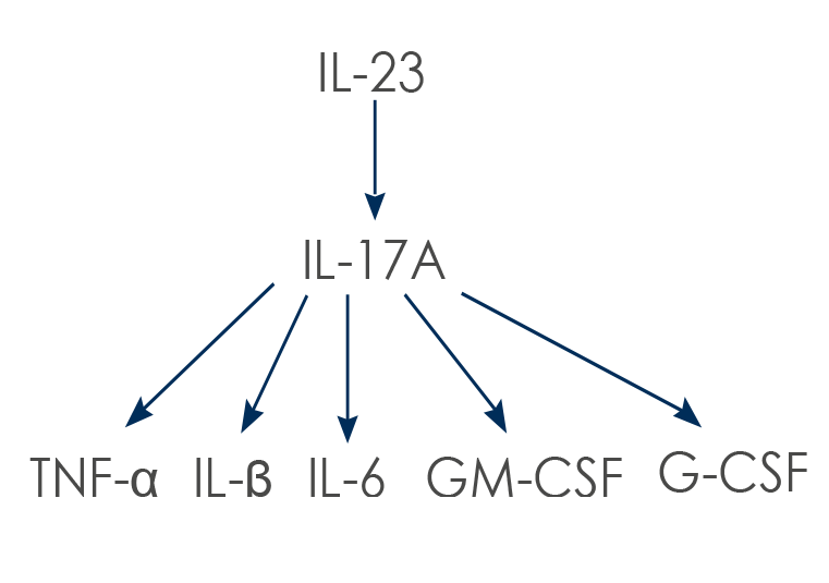
Th17 lymphocytes are stimulated by IL-23; the resulting production of IL-17 increases the synthesis of other cytokines and the migration of another type of immune cells, the neutrophils.
IL-17A and IL-17F (which has a structure very similar to that of IL-17A, with which it shares the same receptors) also act on other cells (such as macrophages, fibroblasts, osteoblasts, chondrocytes and endothelial cells). Their action leads to the synthesis of other pro-inflammatory cytokines: Tumor Necrosis Factor α (TNF-α), IL-1β, IL-6, Granulocyte-Macrophage Colony-Stimulating Factor (GM-CSF) and granulocyte colony-stimulating factor (G-CSF).
Members of the IL-17 family play a key role against infections by bacteria and fungi. However, their excessive activity has been associated with several inflammatory diseases, such as psoriasis, psoriatic arthritis, rheumatoid arthritis and ankylosing spondylitis. By stimulating the expression of enzymes able to degrade cartilage, IL-17 contributes to its destruction, and by activating osteoclasts (the cells that break down bone) it promotes the degradation of bone tissue. It has also been shown that Th17 lymphocytes producing IL-17A are involved in vascular inflammation as well; more generally, the inflammatory molecules produced by Th17 lymphocytes favor chronic inflammation.
During this phenomenon, stimulation by IL-23 increases the pathogenicity of Th17 lymphocytes, leading to the production of both IL-17 and other cytokines, such as interferon γ (IFN-γ), IL-22 and GM-CSF. Often, when acting alone, IL-17 is not very active; however, it can act in synergy with these and other molecules involved in inflammatory processes, such as TNF-α and IL-1β, leading to an increase in the production of other mediators of inflammation, such as IL-6 and IL-8.
IL-17 can, for example, stabilize the mRNA expression activated by TNF-α and induce the expression of the receptor for this cytokine, amplifying the responses triggered by its presence. IL-17A and IL-17F can also induce the expression of pro-inflammatory genes in synergy with molecules such as IL-1, IL-6, G-CSF and matrix metalloproteinases.
In vascular inflammation too, IL-17A amplifies the inflammatory response induced by pre-existing damage, sustaining rather than inducing inflammation. In endothelial cells (the epithelium lining the inside of blood vessels) the combined action of IL-17A and TNF-α induces a pro-inflammatory, procoagulant and prothrombotic state. IL-17A sustains the hyper-reactivity of platelets, amplifies it and takes part in angiogenesis, which is essential in chronic inflammation and in the tissue remodeling associated with autoimmune diseases. Finally, in acute coronary syndrome this cytokine favors platelet function, and in acute myocardial infarction it is associated with the development of thrombi.
Given its actions, IL-17 is considered an interesting therapeutic target. Its inhibition (and that of its receptor) has proved to be an effective approach in the treatment of psoriasis, and several molecules that act by blocking the very processes in which IL-17 is involved are currently under study. Both preclinical and clinical-phase studies have provided solid evidence for the validity of approaches aimed at targeting IL-17 to address inflammatory or autoimmune diseases.
C-reactive protein (CRP) too is considered a marker of inflammation. Its baseline levels vary quite a lot from one individual to another, and this interindividual variability is 35-40% based on hereditary factors. The increases associated with inflammatory processes leave no room for doubt, however, as its levels can multiply by up to a thousand times; in the presence of severe tissue damage, including damage that can be induced by the progression of a tumor, they rise from about 1 μg/ml to over 500 μg/ml within only 24-72 hours.
CRP is produced mainly by liver cells. The stimulus that leads to its synthesis is the rise of pro-inflammatory cytokines such as interleukin (IL) 6, Tumor Necrosis Factor α (TNF-α) and IL-1; in particular, IL-6 is necessary, although not sufficient, to induce the expression of CRP. In the absence of inflammation the endoplasmic reticulum of hepatocytes releases it slowly; its secretion, however, rises rapidly following the rise in the levels of inflammatory cytokines.
Besides hepatocytes, CRP can also be synthesized by smooth muscle cells, endothelial cells, macrophages, lymphocytes and adipocytes. Once the inflammatory stimulus has been removed, its levels should fall, decreasing exponentially within 18-20 hours.
CRP levels have long been used as a marker of infections and of cardiovascular events. In particular, high serum CRP concentrations are a strong independent indicator of cardiovascular disease in otherwise asymptomatic individuals. They have also been associated with prognosis in atherosclerosis, congestive heart failure, atrial fibrillation, myocarditis, aortic valve problems and heart transplantation.
But there is more: CRP expression is also elevated in other disorders associated with inflammation, for example rheumatoid arthritis. What is more, its baseline levels can change because of many other factors, including age, smoking, weight, blood lipid levels, blood pressure and oral hormone replacement therapy. When it is the presence of a disease that alters them, there are very few cases in which they can be lowered by relying on drugs that do not act on the underlying condition.
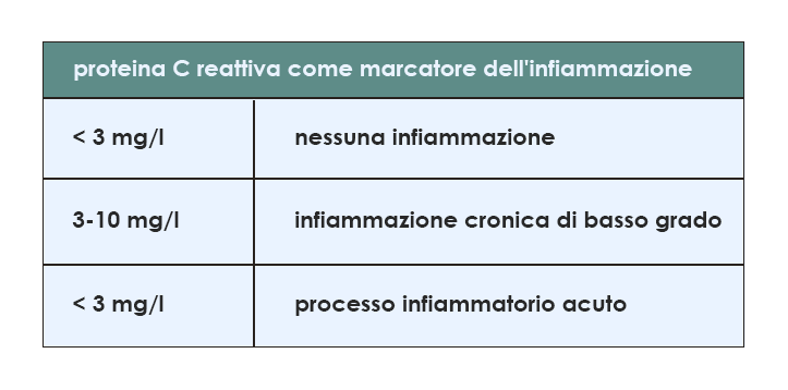
In the association with cardiovascular risk, the rise in CRP is much smaller than the one that can be measured in response to acute inflammation; the threshold level above which the risk is considered significant is set at 3 mg/l. More generally, when used as indicators of the presence of low-grade chronic inflammation, CRP levels are considered elevated if they are between 3 and 10 mg/l, concentrations present in a not negligible share of the population of the Western world (for example, as much as 30% of the inhabitants of North America). Above 10 mg/l, an acute inflammatory process is suspected instead.
CRP is not only a marker, however; rather, this protein plays an active role in inflammatory processes. Some data suggest, for example, that it contributes to the inflammatory response by binding to damaged cell membranes and interacting with the proteins of the complement system (a key component of innate immune defenses), especially in atherosclerotic lesions. There it induces the expression of genes directly involved in the recruitment and adhesion of immune cells; its involvement has also been observed in the mechanisms by which macrophages take up low-density lipoproteins (the form of cholesterol associated with increased cardiovascular risk).
CRP can also stimulate the production of cytokines that favor cell death by apoptosis (IL-1β and TNF-α) and of reactive oxygen species. Apoptosis can also occur within atherosclerotic plaques, favoring their rupture, and CRP can induce it in the smooth muscle cells associated with the coronary arteries.
Finally, in experiments carried out in vitro, CRP significantly reduced angiogenesis, cell migration and the formation of capillary-like structures by attenuating the production of nitric oxide (NO). And it is again in a process associated with the reduction of nitric oxide that CRP promotes atherosclerosis through vasoconstriction, white blood cell adhesion and inflammation. What is more, CRP also influences the levels of IL-6, IL-8 and monocyte chemoattractant protein-1 (MCP-1), a cytokine involved in the migration and infiltration of white blood cells.
Selective removal of cytokines
Keeping the levels of inflammation mediators below the alert limits can help reduce the risk factors associated with the development of diseases linked to inflammation. Bioscience Institute offers an integrated system for the reading and selective removal of excess cytokines in the blood of healthy people who have no symptoms of chronic disease or genetic alterations that precede the development of cancer, but in whom it is possible to identify, or to reduce the risk of, physiological or pre-pathological conditions that predispose to the appearance of these alterations and symptoms.
Through the periodic monitoring of the levels of cytokines in the blood it is possible to follow the rise of one or more inflammation mediators and remove them through an extracorporeal system for blood purification. Cytokine levels are measured again after the purified blood is reinfused, and the operating cycle is repeated until the measured values fall below the attention threshold.
Blood sampling and cytokine removal can take place as a single continuous process or separately. In the second case, if the cytokine levels measured after reinfusion of the purified blood are still too high, a new cycle of sampling and purification is carried out.
After a removal cycle is completed, the next analysis is scheduled based on the cytokine levels measured at the start, the individual's history and other factors set by experts in the field.
Scientific content reviewed by the Bioscience Institute Scientific Board. · Last updated
The information on this page is for general education and does not replace a consultation with a physician.